
Bubble Echocardiogram Interpretation
Dense opacification of the right atrium interatrial sep-tal shift toward the left atrium and bubbles seen crossing septum. If youre a patient interpreting your echo results can give you peace of mind about the next steps towards treatment.
Bubble Contrast Echocardiography Bubble Echocardiogram Test Llc
This helps when it comes to interpreting the echocardiogram reads or echo reads.

Bubble echocardiogram interpretation. A saltwater solution called saline is mixed with a small amount of air to create tiny bubbles and then injected into your vein. Both echo chambers and filter bubbles relate to the ways individuals are exposed to content devoid of clashing opinions and colloquially might be used interchangeably. Consultants who interpret TTE endeavour to provide accu-rate useful reports to colleagues who order these tests.
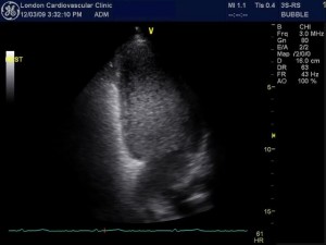
2D echocardiogram api-cal four-chamber view after agitated saline bubble injec-tion. In these select instances our clinic is able to inject a special non-iodinated contrast agent into an IV in your arm in order to improve image quality and our ability to interpret the study correctly. Lungs will filter out all of the saline bubbles or allow for very slow passage of the bubbles to the left side of the heart.
The appearance of these microbubbles in the left atrium LA left ventricle LV or aorta commonly referred to as positive contrast echocardiogram is diagnostic of a right-to-left shunt. Or 2 positive study from other forms of abnormal. Often shortness of breath or chest pain leads your cardiologist to order the test.
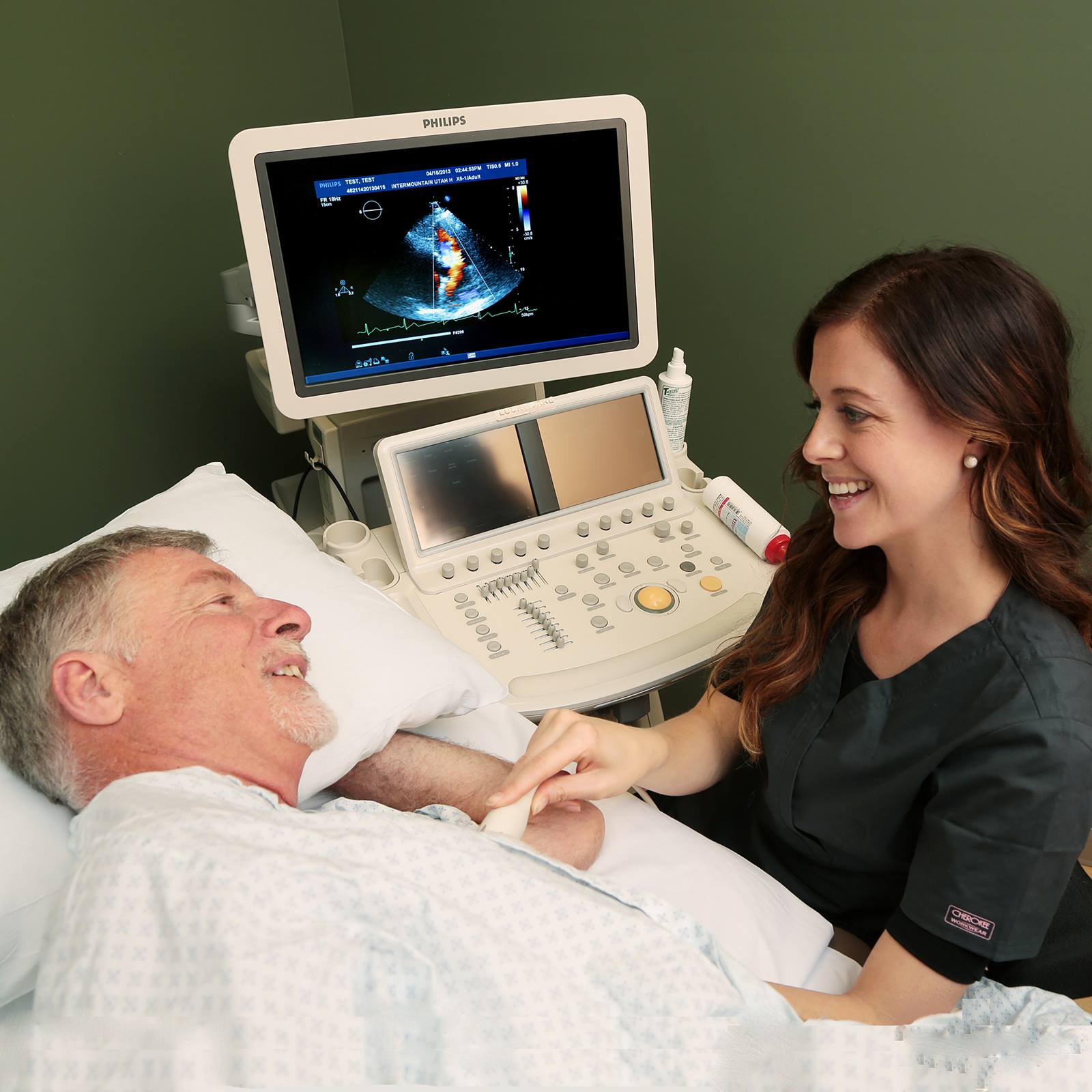
During the bubble study either TTE or TEE. Why am I being asked to come for this test. For the bubble study you will get an intravenous IV line in a vein in your arm.
Use codify for fast cpt code lookup and search. However requires advanced planning. Usually crosses within 1-2 cardiac cycles.
The contrast created by the bubbles allows a left-to-right shunt to be seen as a jet interrupting the opacification of the right atrium. This fluid then circulates up to the right side of your heart and shows up on the echocardiogram image. In a traditional echocardiogram the patients heart is ultrasounded to create a picture of the heart allowing medical professionals to assess the condition of the heart without the need for invasive surgery.
Echocardiograms use high frequency sound waves that are transmitted into the heart to detect its shape size and motion. Video clip is initiated before the injection of the agitated saline at least 2 cardiac cycles before injection Once bubbles start entering the right side we also count how many cycles it takes for bubbles to cross to the left-side. Bubble study can be performed.
The majority of significant holes in your heart are detected in childhood. It is performed to determine you well your heart is pumping blood and whether any structural abnormalities are present. They are normally seen on the right side of the heart before being trapped and absorbed by the pulmonary capillaries so have no route to the left side of the heart.
Having some background knowledge of the purpose of an echocardiogram how the test works and what to look for will help you better understand and interpret echocardiogram results. Your doctor suspects that you may have a hole in your heart. In bubble echocardiogram an iv is injected in the vein of an arm.
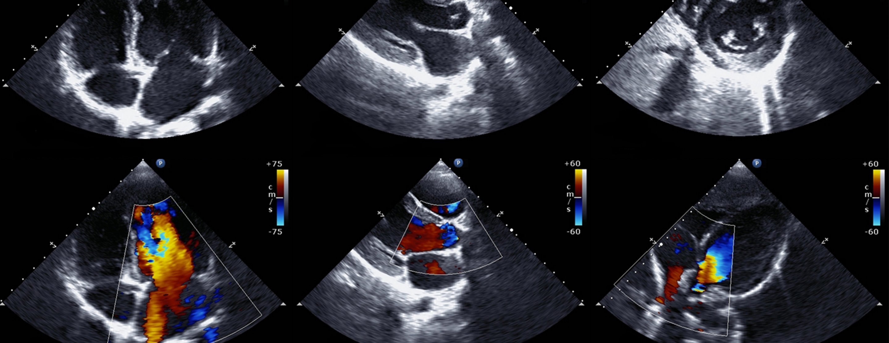
In the setting of pulmonary arteriovenous malformations the timing of right-to-left shunting will vary on the basis of the quantity anatomic location and sizing with prior studies showing the appearance of left atrial bubbles within two to eight cardiac cycles of right atrial opacification. A definite PFO small. If contrast echocardiography is performed by injecting agitated saline solution to a peripheral vein of the upper extremity or neck in patients with Glenn shunt the interpretation of results may be difficult due to 1 poor localization of bubble contrast return from left or right lung.
ContrastBubble echo study Echocardiogram In certain cases regular echocardiograms do not provide adequate image quality for accurate interpretation. A minimum of 4 views would use cpt 72050 and a. A bubble echocardiogram is a procedure which is designed to give a doctor an idea of how well someones heart is functioning.
The passage of bubbles across the septum. Echocardiogram Interpretation and How We Determine Further Imaging Brandon Smith MD. A bubble contrast echocardiogram uses imaging ultrasound combined with an injection of microbubble contrast to help determine additional information.
Stress Echocardiography Echocardiogram performed both before and immediatley after the heart is being stressed Exercise Pharmacological injection. Move to the left and bubbles are seen to cross over. An echocardiogram is a diagnostic test that uses sound waves to create images of your heart.
Few bubbles in a healthy individual may appear due to physiologic intrapulmonary shunt especially in the phase of hypoxia. Its important to know why your doctor prescribed an echocardiogram in the first place. Transesophageal Echocardiography Allows for imaging of intra-cardiac structures.
Ransthoracic echocardiography TTE sometimes called surface echocardiography is a 3basic tool for investigation and follow-up of heart disease. In many US laboratories intravenous bubble contrast is used routinely to outline the LV chamber when the endocardium is poorly outlined. Understanding Echocardiogram Results.
If you have a PAVM the agitated saline bubbles will appear in the left side of the heart after 3-5 beats of the heart and the test would be considered positive for intrapulmonary within the lung shunting blood moving. 60 The specificity of intrapulmonary shunting can be increased by direct visualization of bubbles emanating. Right-to-left shunting occurring primarily with intracardiac lesions such as patent foramen ovale PFO and to a lesser extent via pulmonary arteriovenous malformations PAVM has been associated with a variety of common disease processes12 Thus identification of right-to-left shunting is a frequently requested evaluation in busy echocardiography laboratories.

Pda Echo Findings Cardiac Sonography Medical Photos Nursing School Survival

Echocardiography Ultrasound Physics Vascular Ultrasound Ultrasound

Pin On Echo

Pin On Cardiology
A Typical Apical Four Chamber Echocardiogram Depicting The Four Download Scientific Diagram

Pin By Peggy Gloudemans On Echo Diagnostic Medical Sonography Cardiology Cardiac Sonography

Pin On Serdce Uzi
6 Tips To Improve An Echo Bubble Study

Agitated Saline Contrast Echocardiography In The Identification Of Intra And Extracardiac Shunts Connecting The Dots Journal Of The American Society Of Echocardiography
Echocardiogram
![]()
Transthoracic Echocardiogram Appears To Show A Rigid Membranous Download Scientific Diagram

Echocardiogram Demonstrating Multiple Air Bubbles Yellow Circles In Download Scientific Diagram
Echocardiogram Heart Care Intermountain Healthcare
Echocardiogram Heart Care Intermountain Healthcare
Posting Komentar untuk "Bubble Echocardiogram Interpretation"